- 02/03/2024
- Dr. Ashwini Gaurav
- 0 Comments
- Blog
Normal vs Abnormal Knee X-Ray: Key Differences You Should Know
Knee pain is a common struggle for many Indians, whether it’s due to aging, sports injuries, or the daily strain of commuting. When you visit an orthopedic specialist, the first step toward relief is usually an x ray of the knee. But once you have that black-and-white film in your hand, how do you know if it’s a healthy knee x ray or if something is wrong?
In this blog, we will help you understand the major differences between a normal knee joint xray and an abnormal knee x ray.
This guide is reviewed by Dr. Ashwini Gaurav, the Best Orthopedic Doctor in Patna with over 15 years of experience in joint replacement and bone health.
What is a Knee X-Ray?
An x ray of the knee joint is a quick, painless imaging test that uses a small amount of radiation to create pictures of the bones in your knee. It helps doctors see the knee joint anatomy x ray details, including the femur (thigh bone), tibia (shin bone), and patella (kneecap). While it doesn’t show ligaments or tendons clearly (like an MRI does), it is the “gold standard” for diagnosing fractures and arthritis.
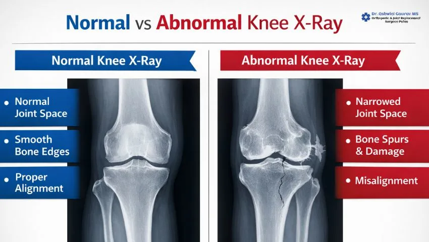
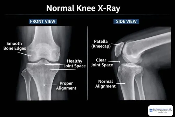
Normal Knee X-Ray: Key Features
In a knee joint normal x ray, everything looks “spacious” and smooth. The normal knee x ray lateral and AP views show:
- Clear, wide gaps between the bones (this is where the cartilage sits).
- Smooth bone edges without any extra lumps or jagged points.
- A perfectly centered kneecap in the normal patella x ray.
- A clean normal leg x ray report with no signs of breaks or thinning of the bone.
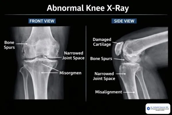
Abnormal Knee X-Ray: Warning Signs
An abnormal knee x ray often looks “crowded” or “rough.” You might see:
- Bones touching each other, indicating the cartilage has worn away.
- Extra bone growths called “spurs” or osteophytes.
- White, cloudy areas indicating high bone density (sclerosis) from friction.
- Dark lines or gaps indicating a knee joint pain x ray finding like a fracture.
Normal vs Abnormal Knee X-Ray: 10 Deep Differences
Understanding the normal vs abnormal knee x ray lateral view and AP view requires looking at these ten major differences:
- Joint Space Width: In a normal knee joint xray, there is a clear, dark gap between the femur and tibia. In an abnormal one, this space narrows significantly, often seen in a bad knee xray.
- Bone Surface Texture: A healthy knee x ray shows smooth, rounded bone ends. Abnormal x-rays show jagged, uneven surfaces where the bone has been damaged.
- Presence of Osteophytes: This is a major difference. A normal xray of knee joint has no extra growths, while an abnormal one shows “bone spurs” sticking out from the edges.
- Bone Density (Sclerosis): In a normal knee x ray, the bone has a consistent grey color. In an abnormal knee x ray, the bone ends often look bright white, showing it is under too much stress.
- Alignment of the Leg: A normal leg xray shows the bones stacking neatly. Abnormal views might show “Bow-legs” or “Knock-knees” (varus or valgus deformity).
- Subchondral Cysts: While a knee normal x ray shows solid bone, an abnormal one may show tiny dark “holes” or cysts just below the joint surface.
- Patellar Position: In a normal patella x ray, the kneecap sits right in the middle. Abnormalities show it tilted to the side or sitting too high/low.
- Loose Bodies: A knee joint normal x ray is clear of debris. An abnormal one might show small white fragments of bone or cartilage floating inside the joint.
- Soft Tissue Shadows: A normal left knee x ray shows very little soft tissue. An abnormal one often shows a “shadow” of swelling or fluid around the joint.
- Surgical Hardware: A natural xray knee normal view shows only bone. If you see metal, it’s an abnormal knee x ray in the sense that it shows a tkr x ray (Total Knee Replacement) or a plate x ray from a past surgery.
Common Conditions Seen in Abnormal Knee X-Rays
- Osteoarthritis (OA): This is the most common reason for a bad knee xray. In osteoarthritis of knee joint x ray, the cartilage wears away, leading to a “bone-on-bone” appearance.
- Rheumatoid Arthritis: Unlike OA, this is inflammatory. The arthritis in knee xray here often shows more “erosions” where the bone looks eaten away.
- Knee Injuries (Fracture/Dislocation): Whether it’s a left knee x ray showing a cracked patella or a right knee x ray with a tibia fracture, the x ray knee is vital for emergency care.
When to Consult a Best Ortho Doctor
If your knee xray images show any of the abnormal signs mentioned above, or if you are experiencing chronic knee joint pain, it is time to see a specialist.
Don’t wait for the pain to become unbearable. Conditions like OA can be managed much better if caught early. If you have already had surgery, your follow-up knee replacement x rays or tkr xray should be monitored regularly to ensure everything is stable.
Conclusion
A knee xray is a powerful tool for your health. Whether you are looking at a normal left knee x ray or a bad knee normal knee x ray comparison, understanding these basics helps you take charge of your recovery.
If you are in Patna and suffering from knee issues, consult Dr. Ashwini Gaurav. With his 15 years of expertise, he can provide a clear diagnosis of your knee x ray photo and recommend the best treatment—from physiotherapy to total knee replacement xray guided surgeries.